by
Gus Iversen, Editor in Chief | March 30, 2015
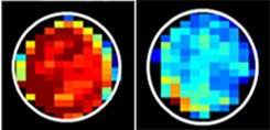
Normal cells (left) have far
more sugar attached to
mucin proteins than do
cancerous cells (right).
Cancerous cells shed sugar from their outer membranes. By visualizing cellular sugar quantities with MRI, researchers from Johns Hopkins University may be paving the way to a no-biopsy-needed cancer diagnosis.
While imaging has always played a crucial role in observing tumors, this would be the first example of visually determining the nature of the cells — malignant or benign — directly and non-invasively.
The researchers published their findings in a report from March 27th in the journal
Nature Communications. Although they have currently only tested their hypothesis on test tube-grown cells and mice, they are hopeful that more clinical benefits can be taken from the study.


Ad Statistics
Times Displayed: 109945
Times Visited: 6642 MIT labs, experts in Multi-Vendor component level repair of: MRI Coils, RF amplifiers, Gradient Amplifiers Contrast Media Injectors. System repairs, sub-assembly repairs, component level repairs, refurbish/calibrate. info@mitlabsusa.com/+1 (305) 470-8013
"We think this is the first time scientists have found a use in imaging cellular slime," said Dr. Jeff Blute, a professor of radiology and radiological science at the Institute for Cell Engineering at Johns Hopkins, and study author.
"As cells become cancerous, some proteins on their outer membranes shed sugar molecules and become less slimy, perhaps because they're crowded closer together. If we tune the MRI to detect sugar attached to a particular protein, we can see the difference between normal and cancerous cells," Blute continued.
The researchers believe that by being able to identify the cancer through its own properties they could theoretically image the entire tumor, something that contrast-aided imaging does not always allow for. Among the practical uses of such imaging capabilities would be chemotherapy-response monitoring, early cancer detection, and guiding biopsies to the most malignant part of a tumor — if not replacing the biopsy entirely.
The next phase in the team's research will involve testing its hypothesis by using MRI sugar imaging to distinguish cancerous tumors from benign masses in mice.


