
How microwaves coupled with machine learning could revolutionize stroke care
October 21, 2024
by Gus Iversen, Editor in Chief
Rapid treatment is critical for minimizing brain damage from stroke, but it’s imperative to first determine if the stroke is hemorrhagic or ischemic. While this is typically done using an MR or CT scan, new research suggests microwaves may offer key advantages for bringing dramatically faster and more affordable diagnosis to one of the world’s most devastating medical emergencies.
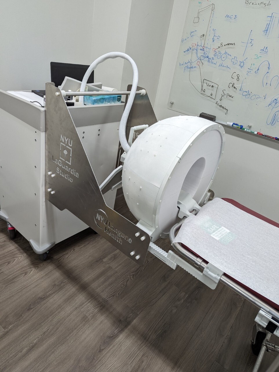
Unlike conventional imaging modalities, the novel 3D-printed system devised by researchers at NYU Langone could weigh about as much as a carry-on bag of luggage, uses less power than a cellphone, and requires no specialized training to operate. It does almost all the work itself, leveraging microwaves and a dedicated set of computational algorithms intended to identify the presence and location of a potential stroke, and generating anatomically realistic MR-like reconstructions of the brain.
HCB News spoke to Dr. Leeor Alon and Dr. Seena Dehkharghani, the co-inventors of the new system. They describe the technology’s inspiration, contrast it with existing tools, and explain why they believe it represents a “new class of imaging and diagnostic instruments” poised to democratize healthcare.
Here is our conversation, lightly edited for clarity and brevity.
 HCB News: Before we get into the technology, why did you and your team set your sights on stroke diagnosis?
Drs. Dehkharghani and Alon: Stroke remains a leading cause of death and disability, with one in four people affected in their lifetime, and disproportionately affects low-income countries and the economically disadvantaged. The resulting economic burden is enormous, with 34% of global total healthcare expenditure attributed to stroke. In the U.S. alone, almost 800,000 individuals are affected every year, and the associated costs are greater than $56 billion.
There are plenty of neurologic disorders that can be serious or even fatal, but none among them carry such rigid guidelines and strict timelines for clinical management and time-to-treatment. Keeping in mind that strokes can relate either to sudden bleeding within the brain (hemorrhagic stroke) or to energetic failure most often resulting from insufficient blood flow beyond a clot (ischemic stroke), treatment planning for a stroke patient generally begins with definitive exclusion of bleeding.
This owes to the fact that the most emergent therapy for ischemic strokes, known as thrombolytics, cannot safely be administered in patients with any sort of hemorrhage. The consequences can be catastrophic; hence thrombolytic treatment is suspended until exclusion of bleeding, which unfortunately cannot presently be performed without CT or MR imaging of the brain, neither of which is routinely available in preclinical environments.
Any delay in imaging translates directly to delays in treatment, and any delay in treatment may translate to astronomical losses in functional brain tissue (potentially on the order of millions of neurons per minute). So, we wanted to devise a way to facilitate diagnosis and subsequent treatment for patients.
HCB News: Much of your work boils down to microwaves. What are microwaves and how do they compare with other forms of diagnostic imaging?
SD and LA: As a specific range within the electromagnetic spectrum, microwaves are ubiquitous and in the modern era enable numerous devices in widespread use, from cellular devices to Bluetooth and other wireless communications.
 Light from other stretches of the electromagnetic spectrum is already very much in use for capturing the inner workings of the body and has been for well over a century, with many readers perhaps familiar with Roentgen’s discovery of X-rays. While highly effective at penetrating the body, X-rays have the drawback of ionization, meaning they can lead to breaking of chemical bonds and formation of reactive free radicals. The incredibly positive contributions of X-rays to human health are undeniable, but certainly concerns such as burn risks or carcinogenesis demand prudent use.
MR is also based on transmission and reception of electromagnetic radiation, but at radio frequencies, and therefore well below the energy thresholds causing ionization. Unfortunately, the cost of procuring, operating, and maintaining these powerful machines is high, making them cost-prohibitive in many parts of the world. While the emergence of "low field" MR scanners is promising, they remain relatively costly and lack the hyper-portability to be deployed in public spaces or true first-responder settings.
In contrast, what we’ve developed is a non-ionizing solution in the form of a helmet that is hyper-portable, estimated at around 10–15 kilograms, and at targeted costs in the tens of thousands of dollars. Our system completes acquisitions in a matter of minutes and operates at under three milliwatts of power (far less than the maximum output power of cell phones). Using custom neural networks developed for characterization of the data obtained by the antenna array within the helmet, the system is designed to function as an intelligent, fully diagnostic stand-alone instrument with tomographic capabilities.
HCB News: Why is this the first I’m hearing about the potential diagnostic value of microwaves?
SD and LA: While the use of microwaves as a probe of tissue properties has been considered for years, it has historically proven difficult to recover, or reconstruct, a true anatomic image in the typical sense. This is because the process of transmitting microwaves, allowing their interactions with tissue, and then estimating the signatures of those interactions based on the data received by the antennas introduces many potential paths that cannot conclusively be localized. The difficulty in localizing those effects presents major challenges when attempting to reconstruct an image of a volume or slice of tissue from such data. Historically, this had been performed across relatively narrow ranges of microwave frequencies.
The key insight into our work is the development of ultra-wideband microwave transmission and reflection across far greater ranges than historically used, leveraging advancements in antenna design and technology.
By increasing the range of interrogation across, for instance, 10 GHz or more, an antenna could probe highly varied interactions in a manner not previously performed and could benefit greatly from the biophysical contrasts across those ranges. Taking the additional step of assembling an array of such antennas, for example in a ring around the object, an unprecedented richness of data could be obtained.
HCB News: So modern computational power was necessary to unlock the utility of microwaves?
SD and LA: We realized early on that data of this sort was well-suited to analysis using large artificial neural networks, in what was still the relative infancy of the field approximately seven years ago. Trained, for instance, from contemporaneous brain MR scans, we felt that we could alleviate the challenge of tomographic microwave reconstruction using a subject’s brain MR as a guide in the learning process, and perhaps even develop direct diagnostic capabilities to circumvent the need for expert interpretations, which are unavailable in many places worldwide.
As previously mentioned, the sort of hardware we’re discussing can be manufactured relatively inexpensively and operates quickly and at very low power. Add it all up, and we foresaw a potential imaging solution that addresses many of the most deeply entrenched problems in medical imaging. The icing on the cake was the possibility that appropriately crafted neural networks could "learn" the dielectric signature of normal and pathologic tissue states, and here the motivation for tuning such a system for detection of strokes became obvious.
HCB News: The artificial neural networks are doing the heavy lifting, but none of this would be possible without the hardware you developed in partnership with NYU’s LaGuardia Studio, right?
SD and LA: That’s correct; With the help of LaGuardia Studio we’ve designed and fabricated a fully-fledged prototype of the helmet. Specifically, the mechanical structure was built to be lightweight, comfortable for patient scanning, and seamlessly interface with all the lab-fabricated electronics necessary for scanning. To date, we’ve scanned a few dozen healthy human subjects, having now documented the potential for anatomically realistic brain imaging in 3D from the system, with true MR-like contrast.
We’ve also developed a dedicated experimental benchtop framework for further advancing the core technology and more intensively studying the properties of the technology in realistic human brain phantoms, which we recently reported in the journal Nature: Communications Engineering.
By 3D printing geometrically varied hemorrhage phantoms with the electrical properties of blood, we can navigate the hemorrhage throughout the head model and rigorously test the performance for detection of hemorrhage, for classification of hemorrhage subtypes, sizes, and shapes, and for precise localization in 3D space. This allows us to optimize the acquisition protocol and neural network in order to streamline it, making it as fast, lean, and as accurate as possible.
HCB News: Where do you see technology like this making the biggest impact?
SD and LA: Any of the challenges described thus far can be thought of as being amplified outside the wealthiest urban areas and are overwhelmingly the burden of the most economically disadvantaged. In less affluent and less industrialized parts of the world there may be literally no access whatsoever for vast stretches across an entire country.
Imagine a system deployable with the ease of something like a defibrillator (forgive the very loose analogy) — a true grab-and-go piece of technology with a footprint small enough and light enough for ubiquitous availability, and at a cost that doesn’t further perpetuate massive imbalances in global health.
What excites us most is the possibility of liberating such diagnostic tools from the requirements of the specialization for operation and interpretation — issues that would remain unaddressed even if we had the ability magically to drop MR scanners into every parcel of land on Earth.
The time is right, maybe even overdue, for us to take advantage of the state of the science in order to transform this segment of healthcare. Biomedical sciences have advanced in extraordinary ways, but we have allowed an untenable and frankly disgraceful disparity in care access to crystallize in the global health landscape. So you can hopefully see that the ethos of our work and the technology is nothing less than a full-bore democratization of advanced and intelligent diagnostic tools.
HCB News: Could microwaves have other diagnostic applications?
SD and LA: We imagine that an expanded array of hardware could be coupled to an expanded armament of algorithms each intended for dedicated purposes beyond just stroke. There are certainly many other high-acuity conditions that would benefit from early detection and diagnosis.
Two that immediately come to mind are traumatic brain injury/concussions and hydrocephalus, a condition in which the fluid spaces of the brain expand to dangerous levels. Here again, many such conditions concentrate in certain economically disadvantaged areas of the world, where a lack of imaging is already a pervasive concern.
We’ve generally preferred not to delimit any strict set of tasks because we see boundless opportunities for what we believe is a new class of imaging and diagnostic instruments.
HCB News: What are the next steps for bringing this scanner to healthcare providers?
SD and LA: We’re at the precipice of clinical subject scanning in patients with neurologic disease, which we hope to initiate with the incredible support offered by NYU Langone in the coming weeks or months.
Larger-scale progress is going to require that we upscale engineering and production of the instrumentation, which will involve some further fine-tuning once we arrive at a design freeze. We’re optimistic that we can rapidly upscale production, given our level of control over the design and engineering from stem to stern.
We’re hoping to secure the necessary funding from varying sources so that we can see this reach fruition, and hopefully after a strong showing in the clinical trial domain, we would naturally apply our efforts fully toward regulatory approval and full commercialization. There’s a certain eagerness and excitement that comes with sitting on something that feels revolutionary and disruptive.
Unlike conventional imaging modalities, the novel 3D-printed system devised by researchers at NYU Langone could weigh about as much as a carry-on bag of luggage, uses less power than a cellphone, and requires no specialized training to operate. It does almost all the work itself, leveraging microwaves and a dedicated set of computational algorithms intended to identify the presence and location of a potential stroke, and generating anatomically realistic MR-like reconstructions of the brain.
HCB News spoke to Dr. Leeor Alon and Dr. Seena Dehkharghani, the co-inventors of the new system. They describe the technology’s inspiration, contrast it with existing tools, and explain why they believe it represents a “new class of imaging and diagnostic instruments” poised to democratize healthcare.
Here is our conversation, lightly edited for clarity and brevity.
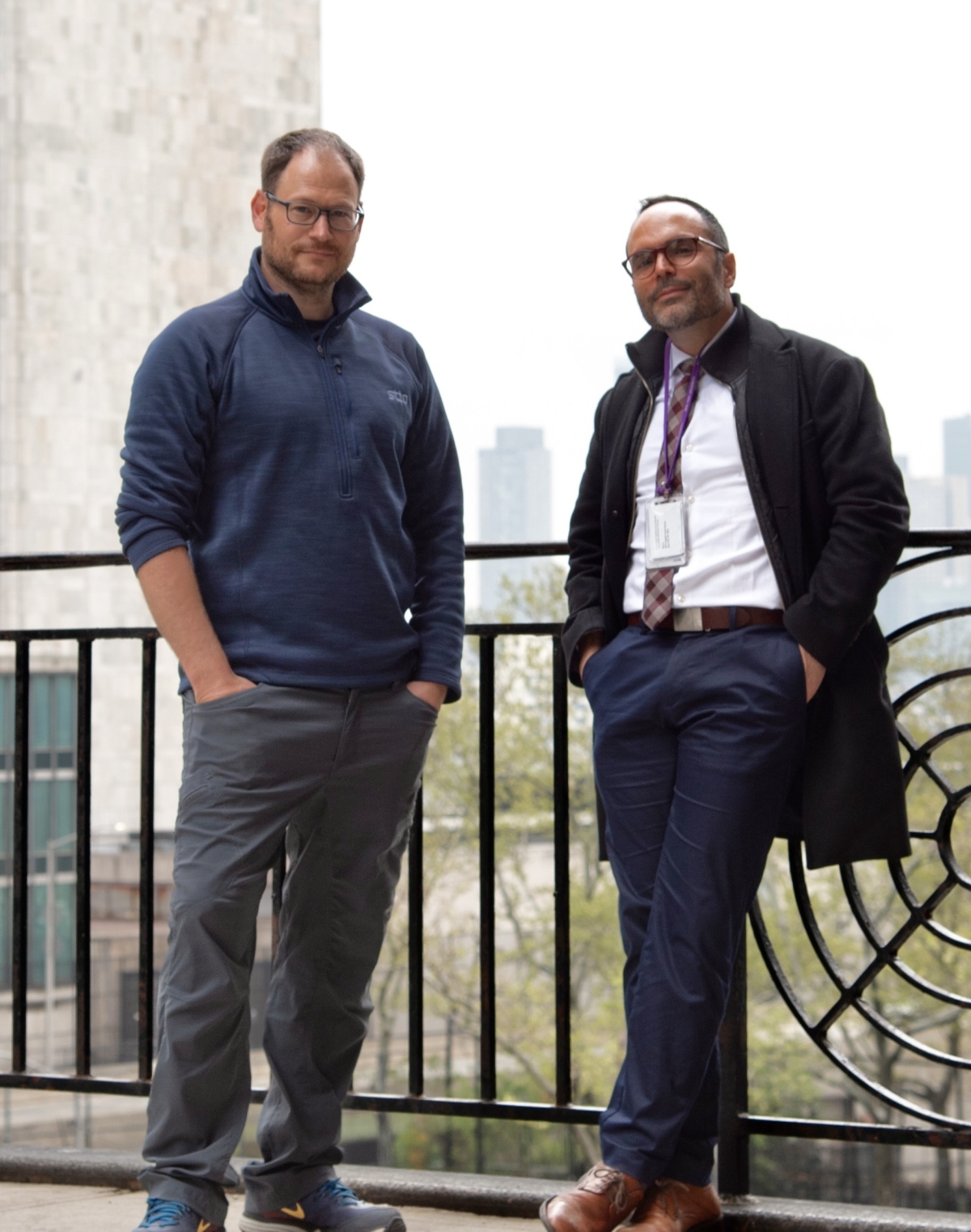
Dr. Leeor Alon (L) and Dr. Seena Dehkharghani (Credit: Pawel Slabiak, NYU Langone Health)
Drs. Dehkharghani and Alon: Stroke remains a leading cause of death and disability, with one in four people affected in their lifetime, and disproportionately affects low-income countries and the economically disadvantaged. The resulting economic burden is enormous, with 34% of global total healthcare expenditure attributed to stroke. In the U.S. alone, almost 800,000 individuals are affected every year, and the associated costs are greater than $56 billion.
There are plenty of neurologic disorders that can be serious or even fatal, but none among them carry such rigid guidelines and strict timelines for clinical management and time-to-treatment. Keeping in mind that strokes can relate either to sudden bleeding within the brain (hemorrhagic stroke) or to energetic failure most often resulting from insufficient blood flow beyond a clot (ischemic stroke), treatment planning for a stroke patient generally begins with definitive exclusion of bleeding.
This owes to the fact that the most emergent therapy for ischemic strokes, known as thrombolytics, cannot safely be administered in patients with any sort of hemorrhage. The consequences can be catastrophic; hence thrombolytic treatment is suspended until exclusion of bleeding, which unfortunately cannot presently be performed without CT or MR imaging of the brain, neither of which is routinely available in preclinical environments.
Any delay in imaging translates directly to delays in treatment, and any delay in treatment may translate to astronomical losses in functional brain tissue (potentially on the order of millions of neurons per minute). So, we wanted to devise a way to facilitate diagnosis and subsequent treatment for patients.
HCB News: Much of your work boils down to microwaves. What are microwaves and how do they compare with other forms of diagnostic imaging?
SD and LA: As a specific range within the electromagnetic spectrum, microwaves are ubiquitous and in the modern era enable numerous devices in widespread use, from cellular devices to Bluetooth and other wireless communications.
MR is also based on transmission and reception of electromagnetic radiation, but at radio frequencies, and therefore well below the energy thresholds causing ionization. Unfortunately, the cost of procuring, operating, and maintaining these powerful machines is high, making them cost-prohibitive in many parts of the world. While the emergence of "low field" MR scanners is promising, they remain relatively costly and lack the hyper-portability to be deployed in public spaces or true first-responder settings.
In contrast, what we’ve developed is a non-ionizing solution in the form of a helmet that is hyper-portable, estimated at around 10–15 kilograms, and at targeted costs in the tens of thousands of dollars. Our system completes acquisitions in a matter of minutes and operates at under three milliwatts of power (far less than the maximum output power of cell phones). Using custom neural networks developed for characterization of the data obtained by the antenna array within the helmet, the system is designed to function as an intelligent, fully diagnostic stand-alone instrument with tomographic capabilities.
HCB News: Why is this the first I’m hearing about the potential diagnostic value of microwaves?
SD and LA: While the use of microwaves as a probe of tissue properties has been considered for years, it has historically proven difficult to recover, or reconstruct, a true anatomic image in the typical sense. This is because the process of transmitting microwaves, allowing their interactions with tissue, and then estimating the signatures of those interactions based on the data received by the antennas introduces many potential paths that cannot conclusively be localized. The difficulty in localizing those effects presents major challenges when attempting to reconstruct an image of a volume or slice of tissue from such data. Historically, this had been performed across relatively narrow ranges of microwave frequencies.
The key insight into our work is the development of ultra-wideband microwave transmission and reflection across far greater ranges than historically used, leveraging advancements in antenna design and technology.
By increasing the range of interrogation across, for instance, 10 GHz or more, an antenna could probe highly varied interactions in a manner not previously performed and could benefit greatly from the biophysical contrasts across those ranges. Taking the additional step of assembling an array of such antennas, for example in a ring around the object, an unprecedented richness of data could be obtained.
HCB News: So modern computational power was necessary to unlock the utility of microwaves?
SD and LA: We realized early on that data of this sort was well-suited to analysis using large artificial neural networks, in what was still the relative infancy of the field approximately seven years ago. Trained, for instance, from contemporaneous brain MR scans, we felt that we could alleviate the challenge of tomographic microwave reconstruction using a subject’s brain MR as a guide in the learning process, and perhaps even develop direct diagnostic capabilities to circumvent the need for expert interpretations, which are unavailable in many places worldwide.
As previously mentioned, the sort of hardware we’re discussing can be manufactured relatively inexpensively and operates quickly and at very low power. Add it all up, and we foresaw a potential imaging solution that addresses many of the most deeply entrenched problems in medical imaging. The icing on the cake was the possibility that appropriately crafted neural networks could "learn" the dielectric signature of normal and pathologic tissue states, and here the motivation for tuning such a system for detection of strokes became obvious.
HCB News: The artificial neural networks are doing the heavy lifting, but none of this would be possible without the hardware you developed in partnership with NYU’s LaGuardia Studio, right?
SD and LA: That’s correct; With the help of LaGuardia Studio we’ve designed and fabricated a fully-fledged prototype of the helmet. Specifically, the mechanical structure was built to be lightweight, comfortable for patient scanning, and seamlessly interface with all the lab-fabricated electronics necessary for scanning. To date, we’ve scanned a few dozen healthy human subjects, having now documented the potential for anatomically realistic brain imaging in 3D from the system, with true MR-like contrast.
We’ve also developed a dedicated experimental benchtop framework for further advancing the core technology and more intensively studying the properties of the technology in realistic human brain phantoms, which we recently reported in the journal Nature: Communications Engineering.
By 3D printing geometrically varied hemorrhage phantoms with the electrical properties of blood, we can navigate the hemorrhage throughout the head model and rigorously test the performance for detection of hemorrhage, for classification of hemorrhage subtypes, sizes, and shapes, and for precise localization in 3D space. This allows us to optimize the acquisition protocol and neural network in order to streamline it, making it as fast, lean, and as accurate as possible.
HCB News: Where do you see technology like this making the biggest impact?
SD and LA: Any of the challenges described thus far can be thought of as being amplified outside the wealthiest urban areas and are overwhelmingly the burden of the most economically disadvantaged. In less affluent and less industrialized parts of the world there may be literally no access whatsoever for vast stretches across an entire country.
Imagine a system deployable with the ease of something like a defibrillator (forgive the very loose analogy) — a true grab-and-go piece of technology with a footprint small enough and light enough for ubiquitous availability, and at a cost that doesn’t further perpetuate massive imbalances in global health.
What excites us most is the possibility of liberating such diagnostic tools from the requirements of the specialization for operation and interpretation — issues that would remain unaddressed even if we had the ability magically to drop MR scanners into every parcel of land on Earth.
The time is right, maybe even overdue, for us to take advantage of the state of the science in order to transform this segment of healthcare. Biomedical sciences have advanced in extraordinary ways, but we have allowed an untenable and frankly disgraceful disparity in care access to crystallize in the global health landscape. So you can hopefully see that the ethos of our work and the technology is nothing less than a full-bore democratization of advanced and intelligent diagnostic tools.
HCB News: Could microwaves have other diagnostic applications?
SD and LA: We imagine that an expanded array of hardware could be coupled to an expanded armament of algorithms each intended for dedicated purposes beyond just stroke. There are certainly many other high-acuity conditions that would benefit from early detection and diagnosis.
Two that immediately come to mind are traumatic brain injury/concussions and hydrocephalus, a condition in which the fluid spaces of the brain expand to dangerous levels. Here again, many such conditions concentrate in certain economically disadvantaged areas of the world, where a lack of imaging is already a pervasive concern.
We’ve generally preferred not to delimit any strict set of tasks because we see boundless opportunities for what we believe is a new class of imaging and diagnostic instruments.
HCB News: What are the next steps for bringing this scanner to healthcare providers?
SD and LA: We’re at the precipice of clinical subject scanning in patients with neurologic disease, which we hope to initiate with the incredible support offered by NYU Langone in the coming weeks or months.
Larger-scale progress is going to require that we upscale engineering and production of the instrumentation, which will involve some further fine-tuning once we arrive at a design freeze. We’re optimistic that we can rapidly upscale production, given our level of control over the design and engineering from stem to stern.
We’re hoping to secure the necessary funding from varying sources so that we can see this reach fruition, and hopefully after a strong showing in the clinical trial domain, we would naturally apply our efforts fully toward regulatory approval and full commercialization. There’s a certain eagerness and excitement that comes with sitting on something that feels revolutionary and disruptive.