From the March 2013 issue of HealthCare Business News magazine
Narrow band imaging, developed by industry heavyweight Olympus, may help physicians detect Barrett’s Esophagus in less time using fewer biopsies. The technology visually enhances superficial layers of the digestive tract by using different color light wavelengths that penetrate the surface at varying depths.
However, the final verdict on whether this technology is a significant advantage over other older methods of GI image enhancement has yet to be determined. Stuart Jackson, general manager of Pro Scope Systems, says the high cost of the procedure may outweigh its purported benefits. “Hospitals pay a high premium for that,” Jackson says, “and it also doesn’t mean they can’t arrive at the same diagnosis using earlier technology.”
Confocal laser microendoscopy is another promising innovation that rapidly speeds up diagnosis: it allows physicians to evaluate tissue on a cellular level in real time—a close-up evaluation that would only otherwise be available through a biopsy. The technology thus improves the patient experience by helping physicians determine a course of treatment on the spot without having to wait for lab results.
“In the past, we took a biopsy, we took to the lab, and had to have the patient sweat it out three or four days more. Here we now have the ability in real time to see if it’s cancer. That’s a major advance in endoscopic imaging,” says Dr. V. Raman Muthusamy, endoscopy director of the UCLA Center for Esophageal Disorders and associate professor of medicine of the digestive diseases division at UCLA. Confocal laser microendoscopy has been available in the U.S. for the last five or six years in most major academic centers.
Among nontraditional endoscopic devices, magnetically controlled pill capsules also show great promise, according to experts. Developed by Olympus and Siemens, this capsule takes pictures of the patient’s stomach. “They’re almost like a drone in your body,” says Muthusamy. Initial trials look promising, with patients giving the device high marks of approval—93 percent thought the examination was comfortable. Exploratory devices like these will eventually eliminate unnecessary invasive procedures, experts say, allowing them to be reserved as last resort procedures for at-risk patients that need more in-depth examination.
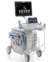
But the technology with the most potential for innovative applications—at least for now—is endoscopic ultrasound. In the procedure, the physician lowers an echoendoscope, an endoscope with ultrasound functionality, down the patient’s GI tract toward the area of interest. Guided by this ultrasound image, the physician inserts a needle directly into the targeted area to take a tissue biopsy. This represents a big leap from other diagnostic imaging techniques such as CT or MRI because physicians are able to obtain a biopsy during the imaging exam and prescribe treatment more quickly. The procedure has already become the gold standard for pancreatic cancer diagnosis.