Rental Only!!!
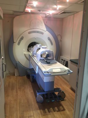
Mobile GE 1.5T HDxt 23x 16-Channel MRI Scanner
It provides 16-channel acquisition capability and HDxt gradients as well as a redesigned user interface. The result is an MR system capable of generating high-definition images in even the most challenging cases. This includes a new operator workspace featuring a wide-screen LCD monitor that hosts a single-screen user-interface and a new host workstation featuring dual-CPUs running a Linux operating system. The advantages include rapid prescription and downloading for greater productivity. The acquisition hardware features a 16-channel receiver architecture and a Volume reconstruction architecture that utilizes blade computing technology, yielding the fastest, most reliable and expandable image reconstruction hardware in the industry. Additional features include a consolidated RF and systems cabinet to reduce space requirements in the equipment room, and a new magnet bridge.
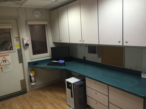
The user interface is at HDxt levels including an LCD wide-screen monitor and keyboard. This flat-panel Liquid Crystal Display (LCD) monitor delivers 1920 x 1200 dot resolution at a refresh rate of 85Hz and an excellent 500:1 contrast ratio using a digital DVI interface -all significant improvements over conventional designs.
Also included in this package:
• Inhance 2.0 Suite
• LAVA Flex
• eDWI
• PROPELLER
• TRICKS
• 16-Channel Head/Neck/Spine Array
• 12-Channel Body Array
• PROBE-PRESS Single Voxel
• PROBE 2D CSI
• PROSE
The Inhance Suite of applications consists of several sequences designed to provide high-resolution images of the vasculature with short-acquisition times and excellent vessel detail. These sequences include: Inhance Inflow IR is a non-contrast-enhanced MR angiography technique that has been developed to image arteries with ability to suppress static background tissue and venous flow. This sequence is based on 3D FIESTA, which improves SNR, as well as produce bright blood images.
Selective inversion pulses are applied over the region of interest to invert arterial, venous, and static tissue. At the null point of the background tissue, an excitation pulse is applied to generate signal. The net result is an angiographic image with excellent background suppression and free of venous contamination. Uniform fat suppression is achieved using a spectrally selective chemical saturation (SPECIAL) technique while respiratory gating compatibility reduces respiratory motion artifacts during free-breathing renal exams.
Inhance 3D Velocity is designed to acquire angiographic images in brain and renal arteries with excellent background suppression in a short scan time. By combining a volumetric 3D phase contrast acquisition with parallel imaging, efficient k-space sampling, and pulse sequence optimization, Inhance 3D Velocity is capable of obtaining the whole neurovascular anatomy in approximately 5-6 minutes. Furthermore, background suppression is improved by the optimized pulse sequence design, resulting in better visualization of small branches. Respiratory triggering is also compatible with Inhance 3D Velocity to enable abdominal angiography, specifically renal arteries. The results are excellent productivity and image quality.
Inhance 3D Deltaflow is a 3D non-contrast enhanced MRA application for peripheral arterial imaging. It is based on cardiac gated 3D fast spin echo and acquires two echoes, one in diastole and the other in systole. Slow arterial flow during diastole results in bright arteries in the diastole images while faster arterial flow during systole results in dark arteries in the systole images. A subtraction of the systole images from diastole images provides arterial-only images with excellent suppression of venous and background signal. Interleaved acquisition and parallel imaging (ASSET) with optimized k-space trajectory helps reduce motion misregistration and improve vessel visualization, respectively. In addition, with the use of partial-Fourier and coronal plane acquisition, the scan time is considerably reduced.
The Inhance 2D Inflow pulse sequence is designed to acquire angiographic images of arteries that follow almost a straight path (i.e, femoral, popliteal, and carotid arteries). Arterial blood flow is foster during the systolic phase and is slower during the diastolic phase. Therefore, Inhance 2D Inflow is designed to acquire data during the systolic phase. It features an optimized spatial saturation gap to improve fat suppression and background suppression. Peripheral Gating is deployed to minimize the pulsatile artifacts Inhance 2D Inflow is compatible with ASSET acceleration to reduce scan time.
LAVA Flex is a 3D FSPGR imaging technique that acquires water only, fat only, in phase and out of phase echoes in one single acquisition that is typically completed in one 20 sec. breath hold. This innovative technique provides excellent homogeneous fat suppression over the entire field of view, including areas that are difficult to image using conventional fat suppression due to magnetic susceptibility effect. As all 4 contrasts are acquired in the same scan, they are perfectly co-registered. The water only contrast differs from a conventional fat suppressed image in that an inversion prep pulse is not applied for fat suppression. In fact, the fat information is removed leaving a water only image that may potentially be used in place of a LAVA type image. LAVA Flex uses ARC, an innovative 2D self-calibrated parallel imaging technique that allows for acceleration in both phase and slice directions for supported coils.
The eDWI (Enhanced Diffusion Weighted Imaging) technique has been designed to provide high signal-to-noise-ratio diffusion images of the liver and brain with short-acquisition time. Its multi-B feature is designed to provide measurement of apparent diffusion coefficient (ADe) map with reduced effect of perfusion. In addition, "3 in 1" technique applies diffusion weighting to all three gradients simultaneously, helping improve sensitivity. Built in tetrahedral feature applies four different diffusion weighing combinations of x, y, and z gradients simultaneously to acquire isotropic diffusion weighted images with high signal to noise ratio and shorter TE. The smart NEX feature significantly reduces the acquisition time. For l.5T, inversion recovery has been deployed to provide robust fat suppression. Enhanced DWI package includes the acquisition sequence and post-processing tools.
PROPELLER (Periodically Rotated Overlapping ParallEl Lines with Enhanced Reconstruction) is a revolutionary data collection technique used in conjunction with the Fast Spin Echo pulse sequence. The name reflects the unique pattern that acquires radial blades of image data rotated in sequence until data acquisition is complete. The redundant data creates images with unusually high contrast-to-noise ratio as well as makes the sequence insensitive to motion artifacts on T2 and T2 FLAIR sequences and insensitive to susceptibility artifacts on DWI sequences. The result is high quality T2 and T2 FLAIR images
of the brain even when the patient fails to remain still, and high quality DWI images in the presence of dental work or surgical hardware. As a result, PROPELLER enables reliable, high quality brain imaging.
TRICKS (Time Resolved Imaging of Contrast KineticS) uses segmented temporal sampling and complex data recombination to accelerate 3D dynamic vascular imaging without compromising spatial detail. TRICKS also uses elliptic centric data collection for optimized contrast resolution and auto-subtraction for optimized background suppression. The result is time course imaging that does not require timing or triggering, provides high temporal and high spatial resolution, and enables the extraction of optimum phases of data. As a result, TRICKS enables reliable, high quality vascular imaging.
1.5T Optima Edition 23 ScanTools Upgrade Pak with PROPELLER 3.0 Upgrade
1.5TOptima Edition 23 ScanTools Upgrade Pak with PROPELLER 3.0 Upgrade
The ScanTools Pak contains 23.0 ScanTools, 23.0 System Software, and English Operator Manual.
The Express Exam and ScanTools 23 1.5T includes a comprehensive suite of workflow features, advanced applications, and parallel imaging capabilities to enable the user to harness the Simply Powerful capabilities of the scanner efficiently and effectively. The patient and technologist Express Exam workflow automates many of the routine tasks that previously required user interaction, thus dramatically reducing the workload for the user and ensuring that consistent and repeatable images are presented for review. Prescription, acquisition, processing and networking steps can be automatically completed throughout the exam. These automated steps can be saved in the Protocol Library to ensure consistent exam workflow for each type of patient.
The automated workflow features of the Express Exam interface includes the Modality Worklist, Protocol Library, AutoStart, AutoScan, AutoVoice, Linking, and Inline Processing that complete much of the work for the user.
Modality worklist: The modality worklist (MWL) provides an automated method of obtaining exam and protocol informat ion for a patient directly from a DICOM Worklist server. For sites with full DICOM connectivity, once a patient has been selected from the MWL, a new session is opened on the host interface and the relevant exam details are highlighted for the user. Additional data fields for patient-sensitive information such as allergies, pre-medication, pregnancy status, and history are provided for greater clarity. The modality worklist provides complete control of the exam protocol prescription. The protocol may be selected well in advance of the patient's arrival at the MR suite thereby simplifying exam preparation and reducing necessary work by the technologist during the time -critical procedure.
Protocol libraries and properties: The 23.0 release provides the user with complete control of protocols for simple prescription, archiving, searching, and sharing. The protocols are organized into two main libraries, a GE Optimized set that are included with the system, and Site Authored. For quick search and selection, each protocol may be archived with independent properties based on patient demographics, anatomy, type of acquisition, or identification number. For commonly used protocols, a favorites flag may be used for quick selection from the Modality Worklist or for sharing across other libraries.
ProtoCopy : Standard on every 1.5Tsystem, the ProtoCopy feature enables a complete exam protocol to be shared with the click of a mouse. The exam protocol can originate from either a library or previously acquired exam. This enables routine archive of protocols for emergency backup and simple management of libraries across multiple systems.
Workflow Manager: Once a protocol has been selected for an exam, it is automatically loaded into the Workflow Manager. The Workflow Manager controls image prescription, acquisition, processing, visualization and networking and may fully automate these steps if requested.
AutoStart: With AutoStart, once the landmark position has been set and the technologist leaves the room the Workflow Manager will automatically start the first acquisition in the exam.
Linking: Linking automates the prescription of images for each series in an exam. Once the targeted anatomical region has been located the Linking feature combines information from a prescribed imaging series to all subsequent series in the Workflow Manager. All series that have been linked may automatically be prescribed (Rx) and no further interaction will be needed by the technologist to initiate the scan. The user has control over which specific parameters can be linked together. Series can have common fields of view, obliquity, slice thickness, anatomical coverage, saturation bands, or shim volumes. Multiple series can be linked together and saved in the Protocol Library or edited in real time. Linking may be used with any anatomy and with any acquisition. Once the first volume is prescribed, all other subsequent series with the same planes, can be automatically prescribed and acquired.
AutoScan: With AutoScan enabled, the Workflow Manager will sequentially go through the list of prescribed series without any user interaction.
AutoVoice: The AutoVoice feature ensures that consistent and repeatable instructions are presented to the patient for each and every exam. User selectable, pre-recorded instructions are presented at defined points in the acquisition. This helps ensure that the patient is in the right position and is fully aware of the next step in the acquisition process. AutoVoice is particularly helpful during breath hold exams. The AutoVoice feature includes instructions in over 14 languages and the user can create and include their own unique voice instructions for local needs.
Inline processing: For certain tasks, the user must accept the results, or complete additional steps prior to saving the images to the database in these cases the data is automatically loaded into the appropriate tool, then the system will await further inst ruction by the user.
Inline viewing: Inline viewing allows the user to conveniently view, compare, and analyze images without having to switch to the Browser. Simply select the series to view from the Workflow Manager and the images are displayed along with standard image display tools. Image comparisons can be easily done by selecting multiple series at a time. The integrated viewer allows the user to seamlessly move between scanning and image viewing.
Image fusion: To better visualize tissue and contrast, multiple images from separate acquisitions can be overlaid on one another. High-resolution anatomical images can be automatically fused with functional data or parametric maps for improved visualization by the user. The data is registered using translation and rotation and distortion correction to ensure accurate fusion. High resolution 20 and 3D data sets
can be fused with reformats, parametric maps, 2D and 3D Spectroscopy maps, plus functional datasets and more.
The automated workflow features of the system can be used for any anatomy and for any sequence. When combining the technology of AutoStart, Linking, Inline Processing, AutoVoice, and the AutoScan features, an entire exam can be completed with just a few actions.
Following is a list of the acquisition pulse sequences and parallel imaging capabilities for HD 23.0. The list is divided into Fast Spin Echo, Gradient Echo, and Echo Planar imaging sequences.
The following sequences are Included for Fast Spin Echo based acquisitions:
Spin Echo: The single echo gold standard for generating T1, proton density and T2 images.
Fast Spin Echo (FSEI. Fast Spin Echo-XL (FSE-XL): Uses a train of spin echoes to reduce total acquisition times and provide high resolution datasets. The sequence provides great flexibility and a large range of imaging options to ensure that quality can be optimized for all anatomy and patient situation.
Fast Recovery Fast Spin Echo (FRFSE): is an extension of the Fast spin Echo sequence and incorporates an additional refocusing pulse and 90 degree excitation at the end of the echo train. This additional forced recovery of the long T1 and T2 spins increases T2 contrast with shorter acquisitions times. The sequence of choice for high-quality, high-speed, and high contrast T2-weighted imaging in neurological, body, orthopedic and pediatric applications.
Single Shot Fast Spin Echo (SSFSE0: An ultra fast scanning technique that permits dataset acquisition within a single RF excitation period. That means it can acquire slices in less than one second, making it an excellent complement to T2-weighted brain and abdominal imaging, as well as MR cholangiopancreatography (MRCP) studies.
FLAIR: T1 and T2 Fluid Attenuated Inversion Recovery (FLAIR) pulse sequences have been designed expressly for neuro applications. FLAIR allows suppression of signal from cerebrospinal flu id (CSF), In addition to this capability; T1 and T2 FLAIR add extraordinary contrast between white and gray matter to T1- and T2-weighted brain and spine imaging.
Double/Triple IR: These pulse sequences are included to allow black-blood imaging for studies of cardiac morphology. Triple IR adds fat suppression to black-blood imaging.
3DFRFSE: A sequence for creating high resolution, three-dimensional T2-weighted images of all anatomies and is especially useful for MR cholangiopancreatography (MRCP) studies. Single-Shot Fast-Spin Echo (SSFSE): An ultra fast technique that permits complete image acquisition following a single RF excitation. It can acquire slices in less than one second, making it an excellent complement to T2-weighted brain and abdominal imaging and MRCP studies.
The following sequences are Included in Gradient Echo based acquisitions:
GRE, FGRE, SPGR, FSPGR: This suite of gradient echo techniques uses short TR and TE times to generate Proton Density-, T1-, T2-, T2* tissue contrast, or a combination thereof, in for less time than conventional spin echo acquisitions. The ultra-short TR and TE times possible with these sequences also ensure the performance needed for state-of-the-art vascular and contrast-enhanced MRA studies.
2D and 3D Dual Echo Gradient Echo: A vital tool for abdominal imaging. This variation on conventional gradient echo provides a pair of images for which the signals from water and fat either are in-phase or out-of-phase. By design, all of the images acquired within a single breath-hold are in perfect registration.
2D and 3D Time of Flight (TOF), 2D-Gated TOF: TOF Imaging and Enhanced 3DTOF Imaging are all ideal for MR angiography. Based on conventional gradient echo scanning, time of flight imaging techniques rely primarily on flow-related enhancements to distinguish moving from stationary spins.
2D Phase Contrast (2DPC), 3D Phase Contrast (3DPC): These techniques demonstrate flow velocities and directional properties in vessels and other moving fluids such as cerebral spinal flu id and aortic flow. These acquisitions provide the data for quantitative flow analysis.
2D MERGE: Multiple Echo Recombined Gradient Echo (MERGE) uses multiple echoes to generate high-resolution images of the C-spine with excellent gray-white matter differentiation. By combining early echoes with high SNR and late echoes with improved contrast, the result is improved cord contrast within the spinal column.
3D MERGE: The 3D MERGE (Multi-Echo Recombined Gradient Echo) sequence has been optimized to generate clear tissue contrast in the cervical spine. By acquiring and summing multiple gradient-echoes at various echo-times, MERGE improves gray-white matter contrast within the cord and provides excellent visualization of the neuroforaminal canals. The high in-plane resolution and thin slices enable excellent image reformats for better tissue visualization from all angles.
COSMIC (Coherent Oscillatory State acquisition for Manipulation if Image Contrast): COSMIC is a 3D imaging technique specifically tailored for Cervical-Spine evaluation. The unique fluid-weighted contrast yields improved visualization of the cervical nerve roots and intervertebral disks. The high resolution images are easily reformatted for better tissue visualization from any orientation.
2D FIESTA: FIESTA (Fast Imaging Employing STeady-state Acquisition) is designed to produce high SNR images extremely rapidly. The technique features an extremely short TR and fully balanced gradients to rephase the transverse magnetization at the end of each TR interval. For very short TR sequences, the signal intensity depends strongly on the ratio T2/T1 and is largely independent of TR. As a result, this pulse sequence accentuates the contrast of spins with a high T2/T1 ratio -- such as CSF, water and fat while suppressing the signal from tissues with low T2/T1 ratio, such as muscle. This property enables high contrast between the myocardium and blood pool.
3D FIESTA: 3D FIESTA (Fast Imaging Employing STeady-state Acquisition) is a technique that uses an extremely short repetition time (TR) between RF pulses such that high-resolution 3D volume images can be acquired rapidly. The 3D FIESTA technique is especially useful for the rapid acquisition of high spatial-resolution images of static structures such as cochlea, internal auditory canal, or joints.
3D FatSat FIESTA: 3D FatSat FIESTA is advanced software designed for imaging of the coronary arteries. The software acquires 3D images using FIESTA (Fast Imaging Employing STeady state Acquisition). Fat suppression is applied to accentuate the coronary arteries. The use of VAST (Variable Sampling in Time) technology greatly shortens breath-holding requirements or allows for higher spatial resolution.
BRAVO-BRAin VOlume Imaging: This IR-prepared 3D Gradient Echo imaging technique affords isotropic, whole-brain coverage with 1x1x1 mm resolution. Coupled with parallel imaging, this sequence produces superior gray white matter contrast in just 2 to 3 minutes.
Brain Volume imaging is a high-resolution 3D gradient echo imaging technique designed to produce heavily T1-weighted isotropic images of the brain in just two to three minutes. BRAVO uses an inversion pulse prior to a train of low flip angle gradient echo acquisitions to reduce scan time and optimize tissue visualization. Bravo is compatible with ARC parallel imaging to minimize scan time and provide whole brain coverage with 1mmx1mmx1mm isotropic resolution.
SPECIAL: Spectral Inversion at Lipids (SPECIAL) is a spectral spatial inversion technique for fat saturation in 3D FGRE pulse sequences.
LAVA: LAVA is a three-dimensional13Dl spoiled gradient echo technique designed specifically to image the liver with unprecedented definition, coverage, and speed in a single breathhold. Excellent fat suppression, through a version of the SPECIAL technique customized for the liver, is one of the reasons for the high definition of anatomical structures. The coverage and speed of LAVA are the result of short TR, innovative use of partial k-space acquisition, and advanced parallel imaging.
For improved tissue contrast, LAVA is compatible with Flex imaging. The LAVA Flex acquisition will provide a water-only, fat-only, in-phase and out of phase data sets in a single acquisition and produce images with significantly reduced chemical shift and susceptibility artifacts.
FastCINE: This pulse sequence is included specifically for studies of cardiac function. Through the use of retrospective gating, it allows full R-R coverage with high multi-phase temporal resolution for excellent visualization of myocardial wall motion.
iDrive Pro: iDrive Pro brings real-time interactive imaging to the MR system, making it easier to generate detailed diagnostic information on just about any anatomy. This includes organs that are subject to motion artifacts, such as spine, heart, diaphragm and GI tract. The iDrive Pro technique allows the user to change scan parameters on the fly, during scanning, to evaluate the results immediately.
SmartPrep: SmartPrep uses a special tracking pulse sequence to monitor the MR signal through a user-prescribed volume to detect the arrival of an injected contrast bolus and to trigger the acquisition one the contrast agent has arrived in the target tissue. Use of SmartPrep provides optimum timing of contrast enhancement.
QuickStep is an automated multi-station acquisition for the evaluation of the vascular tree. This unique application automatically prescribes, acquires, and combines images from multiple stations for fast acquisition and exam completion. To complete the entire exam in as little as 6 minutes, the system will
automatically acquire mask data sets from multiple stations without any user intervention. Secondary images are then acquired at the same independent table positions. The system will automatically subtract the mask images from the secondary dataset and combine the resulting images from the multiple stations into one series. The user only needs to complete a review and approval of the data prior to insertion of images into the database.
The following sequences are included in Echo Planar based acquisitions: An essential tool for any high-throughput site employing advanced techniques. EchoPlanar imaging is what enables the rapid imaging required for such studies as functional brain mapping. And both EchoPlanar and FLAIR EchoPlanar techniques make it easier to generate neuro studies from patients who cannot or will not stay still long enough for conventional techniques.
Diffusion EchoPlanar Imaging: This Diffusion Weighted Single Shot Echo-Planar Imaging (EPI) technique is especially useful for detecting acute and hyper-acute stroke. Its functionality includes Single Shot EPI and FLAIR EPI, Multi-NEX capability, isotropic Diffusion-Weighting imaging and on-line image processing. Diffusion EchoPlanar imaging is the basis for diffusion tensor imaging, sold separately. To enhance body diffusion, Adiabatic SPectral Inversion Recovery (ASPIR) and STIR saturation techniques are supported.
Parallel Imaging Acceleration Approaches: Array Spatial Sensitivity Encoding Technique: ASSET imaging option is an image-based parallel imaging technique used to speed data acquisition. For temporally sensitive acquisitions, ASSET reduces image blurring and motion, enables greater anatomical coverage, and reduces SAR. Parallel imaging acceleration factors up to 3.0 are supported in one dimension depending on the coil selected.
Auto-Calibrating Reconstruction (ARC): Is a GE exclusive self-calibrated parallel imaging technique that eliminates breath-hold mismatch errors by imbedding the calibration data within the scan data. In addition, this unique reconstruction permits small FOV imaging by minimizing focal parallel imaging artifacts from the exam. Supporting both 1D and 2D acceleration, ARC supports high acceleration factors for reduced scan time.
Parallel imaging is supported across all anatomies with acceleration factors that are dependent on the phased-array coils utilized.
IVI: The Interactive Vascular Imaging (IVI) user interface allows operators to quickly remove background from MRA images in order to generate angiographic and maximum intensity (MIP) projections in multiple scan planes. The resulting datasets can be automatically saved as separate series with in a patients exam number, for quick recall in the future.
Multi-Projection Volume Reconstruction (MPVR): MPVR provides quick and easy generation of reformations through any 3D MR data sets.
FuncTool Performance: This package enables advanced MR-image post-processing using a wide range of sophisticated algorithms, including:
• eADCmaps
• Correlation coefficients for mapping of motor strip and visual/auditory stimuli
• NEI (Negative Enhancement Integral)
• MTE (mean time to enhance)
• Positive Enhancement Integral
• Signal Enhancement Ratio
• Maximum Slope Increase
• Maximum Difference Function
• Difference Function
• Diffusion Tensor Post-Processing (requires DTI option)
• 3DCSI Post Processing
MR Pasting: Combine images from separate acquisitions into a single series with MR Pasting. MR Pasting is an image analysis software package that facilitates the display and filming of multiple station MR data sets in the body applications (total spine, total body) as well as peripheral MR angiography data. MR Pasting will automatically register and combine multiple acquisition stations into a single image of covered anatomy.
BrainSTAT software for time course analysis: The BrainSTAT post-processing application automatically generates parametric maps for neuro Blood Flow, Blood Volume, Mean Transit Time, and Time to Peak signal intensity. A Gamma Variant fitting algorithm is used to automatically estimate the arterial input function, and then calculate the quantitative values for the four parametric maps. The maps may be saved in DICOM format and fused with high-resolution anatomic datasets for improved visualization of tissue and anatomy.
R2* Tool: Generate quantitative relaxation maps with the R2 Star (R2*) analysis tools in Functool. With the Express Exam workflow, this feature can automatically generate R2* maps (in units of Hz) and T2* maps (in units of milliseconds) after the multi-echo data has been acquired. The user can have complete control of analysis and may use either the default values to initiate the calculation, or specify specific starting parameter to generate the parametric maps. Input variables for edit include, but are not limited to: number of initial images/echoes to be skipped, lower and upper threshold levels, use of a two-parameter or three-parameter fitting model, confidence level.
The parametric maps may be saved in DICOM format and may overlay high resolution 3D images with Functool Fusion for better tissue visualization. No separate option is necessary to acquire the data; it is included in Express Exam ScanTools.
ConnectPro: The ConnectPro enables the DICOM worklist server class for the Signa operators' console, making it easy to query your HIS/RIS by name, or scheduled date, and to download patient demographics directly to the scanner. The data is automatically loaded into the Express Exam Modality Worklist for simple filtering, editing and prescription of protocols for exam preparation.
ConnectPro may require separate gateway hardware to connect non-DICOM compatible HIS/RIS systems to the MR scanner.
Performed Procedure Step: Performed Procedure Step (PPS) is an important automated connectivity capability - and a key component in film-less and paperless environments. Used in conjunction with the GE PACS broker, it automatically notifies the HIS/RIS and PACS systems of procedure status - in effect, closing the loop on the information gathered from patient arrival through billing. The results: Improved patient care and enhanced productivity.
PROPELLER 3.0 uses an innovative k space filling technique and post processing algorithms to help reduce and correct for motion and minimize magnetic susceptibility artifacts. Radial k space filling pattern causes oversampling of the k space center, generating more SNR and providing excellent tissue contrast. Radial k space filling is inherently less sensitive to motion compared to the Cartesian method.
In addition, a sophisticated motion correction post-processing algorithm is deployed to reduce effects of motion originating from CSF flow, breathing, patient tremor or voluntary movements. PROPELLER 3.0 has been enabled for all anatomies, and T1 FLAIR, T2, T2 FLAIR, DWI as well as PD contrasts in all planes.
1.5T Unified Coil Phantom Kit
Set of phantoms for the 1.5T system that is used on various surface coils to conduct quality assurance testing.
16-Channel 1.5T Head/Neck/Spine (HNS) Array
• The 16-Channel 1.5T Head/Neck/Spine (HNS) Array delivers convenience without compromise. Compatible with systems that have upgraded to the 16-Channel HDxt platform, this 29-element coil serves as a high-resolution brain coil, high-density neuro-vascular array, and a multi-element spine coil in one convenient package. Designed to accommodate multi-dimensional parallel imaging in any scan plane, this coil yields unprecedented imaging speed and superior image quality, thanks in large part to a unique element arrangement that focuses the signal over the anatomy of interest.
12-Channel quadrature Body Array
• The 12-Channel quadrature Body Array with dual connectors is designed for high-definition MR imaging of the chest, abdomen and pelvis on the upgraded 16-channell.5T MR system. This 12-element phased-array coil provides extensive coverage, enabling multi-station anatomical and vascular imaging of the chest-abdomen or abdomen-pelvis without repositioning the coil. The array is optimized for use with ASSET acceleration in enhanced breath-hold imaging procedures.
The MSK Coil 4-Pak includes the following:
• 3-channel Shoulder Array
o The 1.5T 3-channel Shoulder Array offers the increased signal-to-noise characteristic of phased-array technology, along with a unique sleeve design that delivers exceptional joint-imaging capabilities. The coil provides clear definition of the shoulder joint, specifically the head of the humerus, clavicle, acromion, supraspinatus muscle and ligaments. Patient comfort pads and restraining straps are included.
• 8-channel T/R Knee Array
o The 1.5T T/R Knee Array is designed for high definition MR imaging of the knee. This array uses unique hybrid technology using separate birdcage coils for transmit and receive functions. Designed uniquely for GE, the 8-element receive coil delivers 30% to 100% more SNR than the standard extremity coil. The array is compatible with PURE for uniform signal intensity and ASSET for accelerated imaging speed.
• 8-channel Foot and Ankle Array
o The 1.5T compatible foot/ankle coil produces high-resolution images of the foot and ankle by incorporating on 8-channel phased array design in a unique "ski" boot design. The unique coil design has excellent distal coverage and supports multiple foot positions for optimizing studies. Parallel imaging is supported to reduce acquisition times.
• 8-channel Wrist Array
o The 8-Channel Wrist Array generates high definition MR wrist images. The one-piece, ovoid hinged design is optimal for small-FOV imaging and provides 12-cm S/I coverage. The coil can be positioned overhead or at the patient's side, vertically or horizontally. The coil is optimized for ASSET imaging to improve acquisition times.
• Torso Array Coil
• Quad Head Coil
• Neurovascular Array Coil
• Quad Foot/Knee Coil
• Dual Array Package
o 3” Round (2)
o General Purpose Flex Coil (2)
o TMJ Device
o Dual array Adaptor
No Injector
No Camera
AK Coach
Return Policy
Items are sold as-is with no returns or refunds available unless explicitly stated.


 Get a CPS 2-Year Protection Plan starting at $5.99. Sold separately. Learn more